Human Brain Cells Took Over Mouse Cortex — And It Barely Helped
Brain organoids have long promised better disease models than flat petri dishes of cells, but they lack the connections that make a brain a brain. A Stanford team tried something bolder: genetically delete a mouse's cortex, then let human organoid cells fill the vacancy. The graft worked. The payoff did not — at least not yet.
What the Stanford team actually did
The researchers picked a gene active in nearly all cortical cells and used it to trigger deletion of a gene required for chromosome separation during cell division. That killed off most cells destined to form the mature cortex and halved brain volume, yet the mice survived. The team had to cull most other pups so the cortex-free mice got enough nursing, left them with mothers longer, and fed them very high-calorie food. The mice were also immunocompromised to avoid rejecting human cells. Then came the implant: human cortex organoids placed where the mouse cortex would have developed.
The human cells showed up — but didn't organize
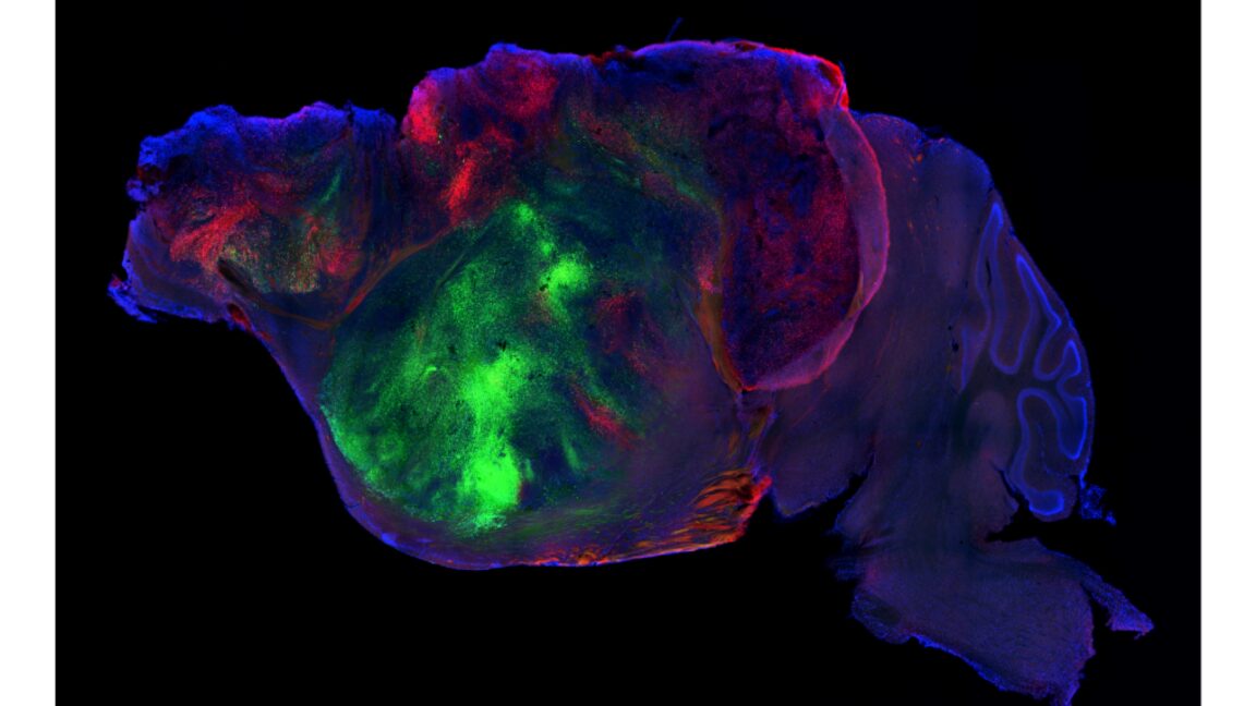
Over 85 percent of implanted animals incorporated the graft, and those human cells went on to make up 92 percent of the cells in the animals' cortex. They formed all major cortical neuron types and even sent processes as far as the spinal cord, with synchronized activity spikes suggesting some coordination. But the structure was missing. Cell types that normally sit in distinct cortical layers ended up near each other without forming those layers at all. Larger-scale organization simply didn't happen — a critical gap if you want to model complex human neural disease.
Behaviorally, the mice landed somewhere in between
In open-field video tracking analyzed by a machine-learning classifier, normal mice, cortex-free mice, and humanized-cortex mice formed three distinct behavioral clusters. Body weight followed the same pattern: cortex-free mice were much lighter, humanized mice fell in between. On a simple maze memory test, cortex-free mice performed at chance, humanized mice beat chance but trailed normal mice. On associative memory, the humanized mice did no better than those with no cortex at all. Fine motor coordination also landed in the middle. Mild gains, no clear cause.
Why this isn't a disease model yet
The team hasn't done the careful anatomical and developmental work needed to say what structures the human cells actually form inside mice, or how much individual variation exists. Without that, the modest behavioral improvements can't be tied to any specific function the human cells provide. One promising signal: the human cells responded differently to brief hypoxia, consistent with normal human cells. But that's far from proving this is a good model for something like ALS. The disorganized connections may never support human neural processing — and without deeper characterization, we won't know.
Key Takeaways
- Stanford researchers genetically deleted most of a mouse's cortex and replaced it with human brain organoids.
- Human cells took over 92 percent of the cortex space and formed all major neuron types, but no layered structure.
- Humanized mice performed between normal and cortex-free mice on memory, weight, and motor tests.
- Associative memory showed no improvement over having no cortex at all.
- Without detailed anatomy and development studies, this isn't yet a usable model for complex neural disease.
Source: Ars Technica • 🇺🇸 San Francisco
Keep Reading